What structure do sex cells have? §35. Sexual reproduction. Formation of germ cells. The concept of parthenogenesis
Helpful information
Sex cells are specialized cells through which the process of sexual reproduction occurs. Female and male germ cells differ from somatic cells (all other cells of the body): they contain half the set of chromosomes. During the process of fertilization, the number of chromosomes is restored. Features of the formation and structure of germ cells ensure their functional specificity.
Female and male reproductive cells: structure
Gametes (sex cells) are characterized by a haploid (single) set of chromosomes. That is, human germ cells contain 23 chromosomes: 22 autosomal and 1 sex chromosome. The types of germ cells (male or female) differ precisely in the sex chromosome: the female germ cell (gamete) contains an X chromosome, the male one contains an X or Y chromosome. During the process of fertilization, the sex of the unborn child depends on the combination of sex chromosomes: XX - female, XY - male.
The structure of germ cells is characterized by incredible structural organization and purposefulness. Male germ cells (spermatozoa), which must be highly mobile in the female reproductive tract, are small cells that lack cytoplasm and consist of a head containing a nucleus with genetic material, and a tail - an organ of movement. Of the cellular elements, they contain only mitochondria, which provide energy for movement, an acrosomal vacuole containing proteolytic enzymes for dissolving the egg membranes, and a proximal centriole. The total length of the sperm is approximately 60 microns, with 55 of them being in the tail.
The acrosomal vacuole of the male germ cell contains the following enzymes:
When sperm emerge from the testicle, they are still morphologically immature; they acquire the ability to fertilize and motility in the vas deferens. In addition, male germ cells contain a number of specific antigens, the inactivation of which also occurs in the vas deferens.
The female reproductive cell (ovum) is a large, immobile cell. It contains a large supply of trophic substances that are necessary for the early development of the embryo. In addition, for the formation of blastomeres (the first generations of embryonic cells), the egg contains a sufficient number of cytoplasmic structures. The human egg is oligolecial, meaning it does not contain much yolk.
A feature of the germ cells of higher placentals, including humans, is that a mature germ cell does not exist in isolation, it is always in close contact with the surrounding somatic cells that create the membrane. The complex of the female reproductive cell with somatic membranes is called the ovarian follicle, or ovosomatic histion.
Formation of germ cells. Fertilization
The process of development of germ cells is very complex and multi-stage. Primary gametes (sex cells) in the embryonic period are laid far from the gonads, and then during development, with a current of moving fluids, they are transferred to the gonad area. Already in the gonads their further formation occurs. During further embryonic development, surrounding cells and tissues do not influence the process of direct formation of gametes, and no acquired human characteristics are inherited.
Formation of female germ cells (ovogenesis)
The formation and maturation of female germ cells occurs in follicles located in the ovarian tissue. Primordial follicles move into the ovarian tissue during embryogenesis. A distinctive feature is that female reproductive cells are formed in large numbers in the ovarian tissue; by the time of birth, their number is about two million. A larger number of cells are resorbed, and by the time of puberty there are approximately 300 thousand oocytes. Female germ cells are formed only in the embryonic period, and before puberty only their final structural formation occurs. That is why absolutely all the negative factors that a woman encounters during her life are reflected in the state of her gametes. The influence of alcohol on reproductive cells at any period of life has an extremely negative effect, and its consequences persist forever. New germ cells in women are not formed during life, only their maturation occurs.
During reproductive age, several follicles mature each menstrual cycle. By the time of ovulation (the period when a mature reproductive cell emerges from the follicle), there is a finally formed dominant follicle. It increases in size, and by the time of ovulation, the cavity with the follicle in the ovary, filled with liquid (graphic vesicle), reaches 2 cm in diameter.
When the follicle matures, the cells surrounding it produce hormones - estrogens. Just before ovulation, their concentration increases significantly, resulting in the release of luteinizing hormone. In this case, the follicle ruptures, and the egg, ready for fertilization, is released into the abdominal cavity, from where it then enters the fallopian tubes.
Development of male germ cells (spermatohegesis)
The male reproductive cell is formed in a completely different way. At the time of birth, the gonads contain rudimentary, unformed male reproductive cells. The process of their final formation begins with puberty. A distinctive feature of the formation of male germ cells is that each cell is formed in approximately 75 days, and not from the moment of birth, like female cells.
The process of sperm formation occurs in the convoluted seminiferous tubules. Spermatogonia (precursors of mature male germ cells) are located on the basement membrane, where they undergo stages of mitotic division. As a result of mitosis, two types of cells are formed. Spermatogonia A retain the ability to further divide by mitosis and give rise to the same cells, while spermatogonia B are evacuated from the membrane and are able to divide only by meiosis. It is after the first meiosis that cells with a single set of chromosomes are formed, which in 75 days finally mature and are ready for fertilization of the egg.
Sex cells: fertilization
The fusion of two sex cells is called fertilization. The fertilization process ends with the formation of a zygote. The sex cells of a woman and a man have a haploid (single) set of chromosomes, and when they merge, the diploid (double) set of chromosomes characteristic of the human body is restored. In this case, the unique genetic information of the maternal and paternal organisms is combined. The formed zygote has the property of typotolerance - it is capable of giving rise to a variety of cells and tissues of the future organism.
The process of fertilization of the egg occurs in the fallopian tube. The sperm, with the help of acrosomal enzymes, destroys the membranes of the egg (corona radiata, zona pellucida), and the process of fusion of its plasma membrane with the membrane of the egg occurs. After this, the head of the sperm penetrates into the cytoplasm of the egg. When the genetic material of the sperm has penetrated the egg, the fertilization process ends, resulting in the formation of a unique new single-cell system, giving rise to a new organism.
When a sperm penetrates the egg, the enzymes released from it modify the membrane in such a way that other sperm can no longer destroy it and penetrate inside the egg. This process takes only a few minutes. Only one sperm takes part in the fertilization process. In extremely rare cases, when two sperm penetrate the egg, a triploid embryo is formed, but it is not viable and dies within a few days.
After fertilization, the zygote stage lasts about 30 hours. Next, crushing begins. This is the process of mitotic division of the zygote, as a result of which the number of its cells increases, but the overall size remains the same. At this stage, the cells are called blastomeres. After 3 days, when all the formed cells are identical in determination and size, the stage of their differentiation begins. On day 5 of development, the embryo is a blastocyst, which consists of approximately 200 cells. A blastocyst is a hollow ball of cells (trophoblast cells) containing embryoblast cells. If a blastocyst contains two embryoblasts, identical twins are formed from such an embryo.
During this entire period, the embryo migrates through the fallopian tube into the uterine cavity. This process occurs under the influence of the movements of the villi on the surface of the fallopian tubes. When the embryo reaches the uterine cavity, implantation occurs. In this case, the blastocyst loses the zona pellucida (this process is called hatching) and, with the help of special processes, sinks into the endometrium. This process is regulated by close chemical and physical connections between the endometrium and blastocyst. Trophoblast cells produce human chorionic gonadotropin, which stimulates the production of progesterone by the cells of the corpus luteum, as a result of which menstruation does not occur.
It is precisely this complexly organized process of development of germ cells that ensures the extraordinary phenomenon in which a new, unique organism is formed from two small cells with a set of unique genetic information - a new person.
The Russian Oocyte Donor Center offers a wide selection of donors to women in need of infertility treatment using donor eggs. Contact you - and we will definitely help you!
Gamete: a germ cell (sperm or egg) containing a haploid set of chromosomes, that is, having one copy of each chromosome.
With the sexual method of reproduction, offspring, as a rule, have two parents. Each parent produces sex cells. Sex cells, or gametes, have a half or haploid set of chromosomes and arise as a result of meiosis. Thus, a gamete (from the Greek gamete - wife, gametes - husband) is a mature reproductive cell containing a haploid set of chromosomes and capable of merging with a similar cell of the opposite sex to form a zygote, and the number of chromosomes becomes diploid. In a diploid set, each chromosome has a paired (homologous) chromosome. One of the homologous chromosomes comes from the father, the other from the mother. The female gamete is called an egg, the male - a sperm. The process of gamete formation has a common name - gametogenesis.
In the embryos of all vertebrates, at an early stage of development, certain cells are isolated as the precursors of future gametes. Such primary germ cells migrate to the developing gonads (ovaries in females, testes in males), where, after a period of mitotic reproduction, they undergo meiosis and differentiate into mature gametes. In germ cells, before meiosis, additional genes are activated that regulate the pairing of homologous chromosomes, recombination and separation of recombined homologous chromosomes in anaphase of the first division.
Eggs develop from primordial germ cells, which, at an early stage of development of the organism, migrate to the ovary and transform there into oogonia. After a period of mitotic reproduction, oogonia become first-order oocytes, which, having entered the first division of meiosis, are delayed in prophase I for a time measured in days or years, depending on the type of organism. During this delay, the oocyte grows and accumulates ribosomes, mRNA and proteins, often using other cells, including surrounding supporting cells, in the process. Further development (maturation of the egg) depends on polypeptide hormones (gonadotropins), which, acting on the auxiliary cells surrounding each oocyte, encourage them to induce the maturation of a small part of the oocytes. These oocytes complete the first meiotic division, forming a small polar body and a large second-order oocyte, which later enters metaphase of the second meiotic division. In many species, the oocyte is delayed at this stage until fertilization initiates the completion of meiosis and the beginning of embryonic development.
The sperm is usually a small and compact cell that is highly specialized for the function of contributing its DNA to the egg. While in many organisms the entire pool of oocytes is formed at an early stage of female development, in males, after the onset of puberty, more and more germ cells enter meiosis, with each first-order spermatocyte giving rise to four mature sperm. Sperm differentiation occurs after meiosis, when the nuclei are haploid. However, since during the mitotic division of mature spermatogonia and spermatocytes cytokinesis is not completed, the descendants of one spermatogonia develop in the form
Sexual reproduction- a method of reproduction in which a new individual usually develops from a zygote resulting from the fusion of two sex cells.
Sexual process. Sexual reproduction is characterized by the presence of a sexual process, during which the germ cells (gametes) come together and their subsequent fusion (fertilization) occurs. Gametes in most organisms are formed with recombined parental chromosomes (remember how meiosis occurs). When gametes fuse, a diploid zygote is formed, from which an organism develops that has inherited a unique combination of genes and characteristics from both parents. Thus, sexual reproduction (as opposed to asexual reproduction) results in a variety of offspring. This increases the ability of organisms to adapt to changing environmental conditions, which is of paramount importance in the evolution of living nature.
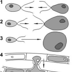
There are two types of sexual process - conjugation and copulation. During conjugation, the contents of two unspecialized cells merge (in some algae And mushrooms) or the exchange of genetic material between individuals (in some bacteria And ciliates). Moreover, in the second case there is no increase in the number of individuals. However, due to the exchange and recombination of genetic material, an increase in the hereditary variability of organisms is ensured.
Copulation (gametogamy) is the fusion of germ cells to form a zygote. In this case, the haploid nuclei of the gametes form the diploid nucleus of the zygote.
The structure of germ cells. In most species of living organisms, two types of germ cells are formed, differing in structure and physiological properties - male (motile sperm or immobile sperm) and female (eggs).
Sperm humans and many animals have a head, neck, middle part and a long flagellum (tail), which serves for active movement (Fig. 79). The head contains a haploid nucleus and a small amount of cytoplasm. At the anterior end of the head there is an ac soma, which is a modified Golgi apparatus. The acrosome contains enzymes that dissolve the membranes of the egg during fertilization. There are two centrioles in the neck, and in the middle part there are mitochondria, which generate the energy necessary for the movement of the flagellum. The tail contains a movable axial filament of the flagellum, built from microtubules.
Sperm can remain viable for a long time outside the body when frozen. This property is widely used in agriculture, in particular when breeding cattle using artificial insemination. The sperm of elite animal breeds is collected and stored in liquid nitrogen, and after thawing, it is used to produce highly productive offspring.
Ovules most often they are motionless and have a spherical shape (Fig. 80). The egg contains a nucleus and cytoplasm with a set of various organelles and a supply of nutrients for the development of the embryo. Therefore, eggs are usually much larger than sperm and somatic cells. For example, the diameter of human eggs reaches 200 microns, while the length of sperm is about 60 microns. The egg cells of animals, the embryonic development of which occurs outside the mother’s body, are very large in size - birds, reptiles, amphibians, fish etc. Yes, Chicken the diameter of the oocyte (an egg without an albumen shell) is more than 30 mm, in some sharks- 50-70 mm, and ostrich- 80 mm.
The eggs are covered with membranes. Based on their origin, shells are divided into primary, secondary and tertiary. The primary membrane of the egg is a derivative of the cytoplasm and is called the vitelline membrane. It is characteristic of the eggs of all animals. Secondary membranes are formed due to the activity of cells that nourish the egg. They are characteristic, for example, of arthropods (chitinous shell). Tertiary membranes arise as a result of the activity of the glands of the genital tract. The tertiary ones include the shell, subshell and albumen membranes of the eggs of birds and reptiles, and the gelatinous membrane of the eggs of amphibians. The membranes of the eggs perform protective functions and ensure the exchange of substances with the environment.
Gametogenesis is the process of formation and development of gametes. In plants, some algae and fungi, the formation of gametes occurs in special organs. For example, in spore plants, female gametes are formed in archegonium, male gametes in antheridia. In most animals, gametogenesis occurs in the gonads.
In nature, there are many species in which the same organism can form both male and female reproductive cells. Such organisms are called hermaphrodites (in Greek mythology, Hermaphroditus is a bisexual creature, the child of the gods Hermes and Aphrodite). Hermaphroditism is common among invertebrate animals ( coelenterates, flat And annelids, mollusks) and in plants.
Formation of germ cells in mammals. The process of formation of male germ cells is called spermatogenesis, and that of females is called oogenesis.
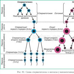
Spermatogenesis occurs in the male gonads - testes. This process is divided into four periods (Fig. 81).
1 . IN breeding season diploid precursors of male gametes — spermatogonia - divide repeatedly by mitosis, which leads to a significant increase in their number. In male mammals (including humans), this process begins at puberty and continues until old age.

2. IN growth period division of spermatogonia stops, and they begin to grow (at the same time, their sizes increase slightly) - first-order spermatocytes are formed.
3. During the maturation period First order spermatocytes divide by meiosis. After the first meiotic division, from each first-order spermatocyte two haploid second-order spermatocytes are formed, after the second - four haploid spermatids.
4 . IN formation period spermatids are transformed into spermatozoa, while the shape of the cell changes, a flagellum, acrosome, etc. are formed.
The duration of spermatogenesis in humans is about 75 days. A huge number of sperm are formed in the testes (testes), for example, in humans, 1 ml of seminal fluid contains up to 100 million.
Oogenesis occurs in the female sex glands - the ovaries - and begins even before birth. In the process of oocyte formation, three periods are distinguished (see Fig. 81).
1. IN breeding season diploid egg precursors — ogonia - divide repeatedly mitotically. In mammals, this process occurs in the embryonic period (before birth). The number of oogonia in the ovaries increases significantly, and then they remain unchanged until puberty.
2. With the onset of puberty, individual oogonia periodically enter into growth period which can last for several months. During this time, their volume increases significantly due to the intake of substances from the surrounding follicular cells and blood. This is how first-order oocytes are formed.
3. Periodically, first-order oocytes enter meiosis. This - period of maturation. During the process of meiosis, daughter cells of different sizes are formed. After the first meiotic division, a large haploid cell is formed - a second-order oocyte - and a small one, called the primary polar body. Ovulation occurs - a second-order oocyte leaves the ovary into the abdominal cavity. It then enters the fallopian tube, where it undergoes a second meiotic division, forming a large egg and a small secondary polar body. The primary polar body, as a rule, also divides into two. All polar bodies subsequently die and are destroyed.
Thus, in contrast to spermatogenesis, where four equal haploid cells are formed during meiosis, during oogenesis one large egg and three small polar bodies develop. The biological meaning of uneven division is to preserve in the egg the maximum amount of nutrients necessary for the future embryo.
1. What are the names of the organs in which the formation of female and male gametes occurs in spore plants? In animals?
Ovaries, antheridia, sporangia, testes, archegonia.
2. How is the structure of the sperm and egg related to the functions performed by these cells?
3. Spermatozoa contain practically no cytoplasm and nutrients, but they need a large amount of energy to move. Where do you think this energy comes from?
4. What is the maximum number of eggs and secondary polar bodies that can be formed in a cat from four first-order oocytes?
5. What processes occurring during oogenesis ensure the accumulation of large amounts of nutrients in eggs?
6. What is the biological meaning of the formation of polar bodies during oogenesis?
7. Compare the processes of spermatogenesis and oogenesis, indicate similarities and differences.
8. The ovaries of a 22-year-old woman with a stable 28-day reproductive cycle contain 42 thousand follicles. Most of them are very small, and only 299 have a diameter greater than 100 microns. In addition, there are 5 corpora lutea in the ovaries and 112 scars remaining from them. At what age did this woman first ovulate? At what age will she most likely stop producing eggs?
- § 1. Content of chemical elements in the body. Macro- and microelements
- § 2. Chemical compounds in living organisms. Inorganic substances
- § 10. History of the discovery of the cell. Creation of cell theory
- § 15. Endoplasmic reticulum. Golgi complex. Lysosomes
Chapter 1. Chemical components of living organisms
Chapter 2. Cell - structural and functional unit of living organisms
Chapter 3. Metabolism and energy conversion in the body
Remember!
Where in the human body do germ cells form?
Eggs - female reproductive gametes are formed in the ovaries, paired organs. Spermatozoa - male reproductive cells are formed in the testes and paired organs.
What set of chromosomes do gametes contain? Why?
A haploid set is a half set of chromosomes, single (odd number), such a set is contained in germ cells (gametes) and is designated n. For example, the haploid set of human chromosomes is n=23. Since when two germ cells are fertilized, the full diploid set of the organism – the zygote – is restored.
Review questions and assignments
1. Compare the structure of male and female reproductive cells. What are their similarities and differences?
Ovules are relatively large, stationary, rounded cells. In some fish, reptiles and birds, they contain a large supply of nutrients in the form of yolk and have sizes from 10 mm to 15 cm. The eggs of mammals, including humans, are much smaller (0.1-0.3 mm) and the yolk is almost do not contain. Spermatozoa are small, motile cells; in humans, their length is only about 60 microns. In different organisms they differ in shape and size, but, as a rule, all sperm have a head, neck and tail, which ensure their mobility. In the head of the sperm there is a nucleus containing chromosomes, and an acrosome - a special vesicle with enzymes necessary for dissolving the egg shell. Mitochondria are concentrated in the neck, which provide the moving sperm with energy; their length is only about 60 microns.
The egg has:
Big sizes
Round shape
The presence of a large amount of yolk (nutrients for the future embryo)
Presence of egg membranes
Sperm have:
Small sizes
Various forms in different mammals
Organ of locomotion (flagella from 1 to several)
Large number of mitochondria
Absence of ribosomes and ER, modified Golgi apparatus.
2. What determines the size of eggs? Explain why mammalian eggs are among the smallest.
From the supply of nutrients. In mammals, development occurs in the womb; its size cannot be large, since the embryo develops in the uterus; the uterus itself is penetrated by blood vessels, which also serve as a source of nutrients and oxygen.
3. What periods are distinguished in the process of development of germ cells?
Stage 1 – reproduction of primary germ cells
Stage 2 – growth of germ cells
Stage 3 – maturation of germ cells
Stage 4 – formation of germ cells (only for spermatogenesis); in oogenesis, at stage 4, the polar body dies off or the formation of egg membranes occurs.
4. Tell us how the maturation period (meiosis) occurs during spermatogenesis; oogenesis.
The third stage is meiosis. Meiosis is a special method of cell division, leading to a halving of the number of chromosomes and the transition of the cell from a diploid to a haploid state. Future gametes at the maturation stage are divided twice. Cells that begin meiosis contain a diploid set of already doubled chromosomes.
The prophase of the first meiotic division (prophase I) is much longer than the prophase of mitosis. At this time, the doubled chromosomes, each of which already consists of two sister chromatids, spiralize and acquire compact sizes. Then the homologous chromosomes are arranged parallel to each other, forming so-called bivalents or tetrads, consisting of two chromosomes (four chromatids). An exchange of corresponding homologous regions (crossing over) can occur between homologous chromosomes, which will lead to recombination of hereditary information and the formation of new combinations of paternal and maternal genes in the chromosomes of future gametes. By the end of prophase I, the nuclear envelope is destroyed.
In metaphase I, homologous chromosomes are arranged in pairs in the form of bivalents, or tetrads, in the equatorial plane of the cell, and spindle threads are attached to their centromeres.
In anaphase I, homologous chromosomes from the bivalent (tetrad) move toward the poles. Consequently, only one of each pair of homologous chromosomes ends up in each of the two resulting cells - the number of chromosomes is halved, and the chromosome set becomes haploid. However, each chromosome still consists of two sister chromatids.
In telophase I, cells are formed that have a haploid set of chromosomes and double the amount of DNA. After a short period of time, the cells begin the second meiotic division, which proceeds as a typical mitosis, but differs in that the cells participating in it are haploid.
In prophase II, the nuclear envelope is destroyed.
In metaphase II, the chromosomes line up in the equatorial plane of the cell, and the spindle threads connect to the centromeres of the chromosomes.
In anaphase II, the centromeres connecting sister chromatids divide, the chromatids become independent daughter chromosomes and move to different poles of the cell.
Telophase II completes the second division of meiosis.
During spermatogenesis at the maturation stage, as a result of meiosis, four identical cells are formed - sperm precursors, which at the formation stage acquire the characteristic appearance of a mature sperm and become motile. Every month, in one of the woman’s ovaries, one of the cells that have stopped dividing continues to develop. As a result of the first division of meiosis, a large cell is formed - the precursor of the egg and a small, so-called polar body, which enter the second division of meiosis. At the metaphase II stage, the precursor of the egg ovulates, that is, leaves the ovary into the abdominal cavity, from where it enters the oviduct. If fusion with the sperm does not occur, the cell that has not completed division dies and is excreted from the body. Polar bodies serve to remove excess genetic material and redistribute nutrients in favor of the egg. Some time after division they die.
6. What is the biological meaning and significance of meiosis?
1) is the main stage of gametogenesis;
2) ensures the transfer of genetic information from organism to organism during sexual reproduction;
3) daughter cells are not genetically identical to the mother and to each other.
And also, the biological significance of meiosis lies in the fact that a decrease in the number of chromosomes is necessary during the formation of germ cells, since during fertilization the nuclei of the gametes fuse. If this reduction did not occur, then in the zygote (and therefore in all cells of the daughter organism) there would be twice as many chromosomes. However, this contradicts the rule of a constant number of chromosomes. Thanks to meiosis, the sex cells are haploid, and upon fertilization, the diploid set of chromosomes is restored in the zygote
Think! Remember!
1. The organism developed from an unfertilized egg. Are its hereditary characteristics an exact copy of the characteristics of the maternal organism?
Yes. This type of reproduction is called parthenogenesis. Parthenogenesis (Parthenogenesis - from the Greek parthenos - girl, virgin + genesis - generation) is a form of sexual reproduction in which the development of an organism occurs from a female reproductive cell (egg) without fertilization by a male one (sperm).
This is sexual, but unisexual reproduction, which arose in the process of the evolution of organisms in dioecious forms. In cases where parthenogenetic species are represented only by females, one of the main biological advantages of parthenogenesis is the acceleration of the rate of reproduction of the species, since all individuals of such species are capable of leaving offspring. If a female develops from fertilized eggs, and a male from unfertilized eggs, parthenogenesis contributes to the regulation of the number and sex ratio (for example, in bees, males - drones - develop parthenogenetically, and from fertilized eggs - females - queens and worker bees).
Parthenogenetically, either an egg that has undergone meiosis and contains a haploid set of chromosomes (n) (generative, haploid, or meiotic parthenogenetic) can develop, or an egg from one of the premeiotic stages of oogenesis with preservation of the chromosome set characteristic of this species - diploid (2n) or polyploid (3n, 4n, 5n rarely 6n, 8n) (ameiotic parthenogenesis). In some forms of parthenogenesis, the fusion of the haploid nucleus of the egg with the haploid nucleus of the directional (polar) body leads to restoration of diploidity (automictic parthenogenesis). The genotype, sex of the parthenogenetic offspring, as well as the preservation or loss of heterozygosity, acquisition of homozygosity, etc., depend on these features of parthenogenesis.
2. Explain why there are two terms for male reproductive cells: sperm (for example, in angiosperms) and sperm.
Spermatozoa are male reproductive cells that have the ability to actively move due to the flagellum. Sperm is a male reproductive cell of plants (gymnosperms, angiosperms), devoid of flagella; moves passively - as a result of the growth of the pollen tube.
These cells differ significantly between men and women. In men, germ cells or sperm have tail-like projections () and are relatively mobile. Female reproductive cells, called eggs, are immobile and much larger than male gametes. When these cells fuse in a process called fertilization, the resulting cell (zygote) contains a mixture of what is inherited from the father and mother. Human sex organs are produced by the organs of the reproductive system - the gonads. produce sex hormones necessary for the growth and development of primary and secondary reproductive organs and structures.
The structure of human germ cells
Male and female reproductive cells differ greatly in size and shape. Male sperm resemble long, mobile projectiles. These are small cells that consist of a head, middle and tail parts. The head contains a cap-like covering called an acrosome. The acrosome contains enzymes that help the sperm cell penetrate the outer membrane of the egg. located in the head of the sperm. The DNA in the nucleus is tightly packed and the cell does not contain much. The middle part contains several mitochondria that provide energy for. The tail consists of a long projection called a flagellum, which aids in cellular locomotion.
A woman's eggs are one of the largest cells in the body and have a round shape. They are produced in the female ovaries and consist of a nucleus, a large cytoplasmic region, a zona pellucida and a corona radiata. The zona pellucida is a membrane covering that surrounds the eggs. It binds sperm cells and helps in fertilization. The corona radiata is the outer protective layer of follicular cells surrounding the zona pellucida.
Formation of germ cells
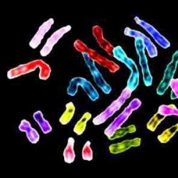
Human germ cells are produced through a two-step process of cell division called. Through a series of sequential events, the replicated genetic material in the parent cell is distributed among the four daughter cells. Since these cells have half the number of the parent cell, they are . Human germ cells contain one set of 23 chromosomes.
There are two stages of meiosis: meiosis I and meiosis II. Before meiosis, chromosomes are replicated and exist in the form. At the end of meiosis I, two are formed. The sister chromatids of each chromosome in the daughter cells are still linked. At the end of meiosis II, sister chromatids and four daughter cells are formed. Each cell contains half the chromosomes of its parent cell.
Meiosis is similar to the process of division of non-sex cells known as mitosis. produces two daughter cells that are genetically identical and contain the same number of chromosomes as the parent cell. These cells are diploid because they contain two sets of chromosomes. Humans have 23 pairs or 46 chromosomes. When germ cells unite during fertilization, the haploid cell becomes a diploid cell.
The production of sperm is known as spermatogenesis. This process occurs continuously inside the male testicles. Hundreds of millions of sperm must be released for this to happen. The vast majority of sperm do not reach the egg. During oogenesis, or egg development, daughter cells divide unevenly in meiosis. This asymmetric cytokinesis results in the formation of one large egg (oocyte) and smaller cells called polar bodies, which degrade and are not fertilized. After meiosis I, the egg is called a secondary oocyte. The secondary oocyte will complete the second stage of meiosis if the fertilization process begins. Once meiosis II is completed, the cell becomes an egg and can fuse with a sperm cell. When fertilization is complete, the combined sperm and egg become a zygote.
Sex chromosomes
Male sperm in humans and other mammals are heterogametic and contain one of two types of sex chromosomes: X or Y. However, female eggs contain only the X chromosome and are therefore homogametic. Sperm of an individual. If a sperm cell containing an X chromosome fertilizes an egg, the resulting zygote will be XX or female. If the sperm cell contains a Y chromosome, then the resulting zygote will be XY or male.


